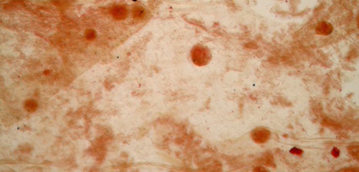
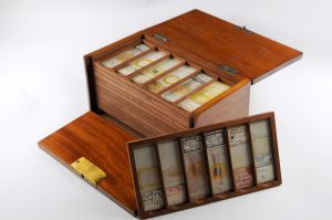
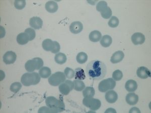
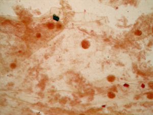
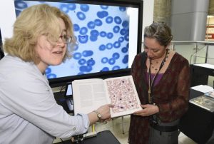
With the great expertise and enthusiasm of Ailie Robinson, Mojca Kristan and staff from the Public Health England Malaria Reference Laboratory and LSHTM Diagnostic Parasitology Laboratory, slides were examined under the microscope, and wonderfully, they were still viable and presented some exciting results. The staining techniques for slides – for example the use of gentian violet – has changed considerably since the 19th century, but oocysts and gametocytes were seen, and the results were photographed under the microscope by Cheryl Whitehorn.
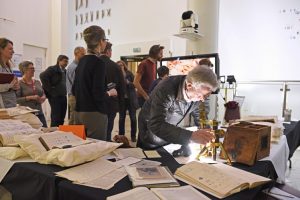
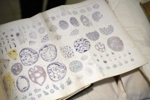
As part of Explore Your Archive week, the resulting images were shown in the Manson foyer, along with an array of rare books on the subject of malaria and a selection of Ross’s archives, including his own early microscopy photographic prints, and his renowned notebook where he meticulously recorded his dissection of the mosquito mid-gut to prove the mosquito as malarial vector.
The event was crowded with enthusiastic students and staff: Myriam Willis, Cheryl and Mojca were on hand throughout the event to explain the slides in detail!
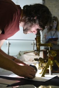
An additional first: a visiting academic, using light from his mobile phone managed to view one of Sir Ronald Ross’s slides through Ross’s very own brass microscope, dating from 1902.
History in the making indeed!
Email archives@lshtm.ac.uk for more information on this project, and for our collections see our catalogue
